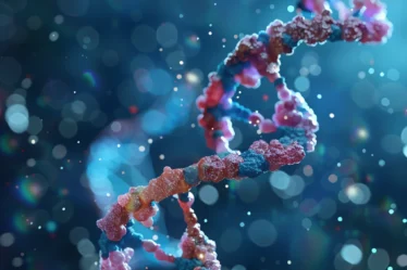
A single human cell holds about two meters of DNA, yet its nucleus measures only around 10 micrometers. This tight packing isn’t random—it’s managed by specific proteins that fold and organize the DNA into structured fibers. Without this organization, the DNA couldn’t fit or function properly inside the nucleus.
This study guide explains how chromatin forms through DNA folding and protein binding. It covers nucleosomes, histone types, euchromatin, heterochromatin, chromatin fiber levels, and structural changes during the cell cycle. You’ll also see how centromeres, telomeres, and chromatin remodeling affect structure and organization in different cell conditions.
Chromatin: Quick Summary
Do you just need the basics? Here’s a simple explanation of what chromatin is and how it works:
🟠 Chromatin is a DNA–protein complex found in eukaryotic cells that helps package long DNA strands into a compact form.
🟠 The nucleosome is the basic unit of chromatin, made of eight histone proteins with DNA wrapped around them.
🟠 Euchromatin is loosely packed and transcriptionally active, while heterochromatin is dense and usually inactive.
🟠 During mitosis, chromatin condenses into chromosomes through a series of folding steps involving fibers and loops.
🟠 Histone modifications and chromatin remodelers regulate DNA accessibility by changing how tightly chromatin is packed.
🟠 Special chromatin structures form at the centromeres and telomeres, supporting chromosome separation and protecting chromosome ends.
Chromatin Structure in Eukaryotic Cells
You can’t fit two meters of DNA into a tiny cell nucleus without folding it carefully. Eukaryotic cells solve this by forming chromatin—DNA tightly wound around proteins. This keeps the DNA organized, stable, and compact enough to fit inside the nucleus.
Chromatin exists in all eukaryotic chromosomes. It contains DNA and about twice its weight in proteins. Most of these proteins are histones. They carry a positive charge and bind easily to the negatively charged DNA, wrapping it into compact units. These units make the DNA shorter and more stable.
In addition to histones, chromatin has many non-histone proteins. They help shape the DNA and control access to different regions when needed. Prokaryotes don’t have chromatin. They package their circular DNA with different proteins into a structure called a genophore, which sits in the nucleoid region.
| Component | Type | Function |
| DNA | Nucleic acid | Genetic code |
| Histones | Basic proteins | Wrap DNA into nucleosomes |
| Non-histone proteins | Structural/enzymatic | Assist folding and access |
Nucleosome Structure and Function
Nucleosome Components and Assembly
A nucleosome forms when DNA wraps around a set of eight histone proteins—two each of H2A, H2B, H3, and H4. This wrapping makes about 1.65 turns and covers 146 base pairs. Short DNA segments called linker DNA connect one nucleosome to the next. Histone H1 binds to the linker and locks the structure into place, forming what’s known as a chromatosome. This setup helps the DNA stay compact but still accessible. Cells can adjust this structure when they need to copy or read certain genes.
Beads-on-a-String Appearance
Nucleosomes line up along the DNA strand like beads, separated by 20 to 60 base pairs of linker DNA. Each unit—core and linker together—adds up to roughly 200 base pairs. This pattern gives chromatin a beads-on-a-string appearance under an electron microscope. Micrococcal nuclease experiments support this: the enzyme cuts exposed linker regions and leaves behind regular-sized DNA fragments. This flexible 10-nanometer fiber is the starting level of DNA packaging and keeps the genome organized without blocking access to important regions.
Higher-Order Chromatin Structures
From Fiber to Chromosome
Once nucleosomes form the 10-nanometer fiber, the chromatin folds into a thicker 30-nanometer fiber. This step shortens the DNA further and adds structure. Scientists propose two main models: the solenoid model, where nucleosomes spiral tightly, and the zig-zag model, where the linker DNA stretches across to alternate sides. Both models explain possible ways to pack DNA, but current X-ray and electron microscopy studies haven’t confirmed a single structure. During mitosis, the 30-nanometer fiber folds into loops and stacks into the dense metaphase chromosome seen under a microscope.
Species Variations in Chromatin Structure
Chromatin doesn’t always follow the same folding rules. Sperm cells pack DNA tightly by swapping histones for protamines. Some protozoa like trypanosomes don’t form visible chromosomes at all. Bird red blood cells and certain specialized cells show other folding patterns. These differences show that chromatin can change based on the cell’s needs and type.
Euchromatin and Heterochromatin
Euchromatin Structure
Euchromatin is loosely packed and easier to access. It contains genes that are often active and is the site where transcription usually takes place. You’ll find it in two main forms: the flexible 10-nanometer fiber and looped sections of the 30-nanometer fiber. This structure allows enzymes and transcription factors to reach the DNA. Under a microscope, euchromatin appears lighter because of its open layout and lower compaction level.
Heterochromatin Structure
Heterochromatin is densely packed and holds long stretches of repetitive DNA. It stays compact and rarely opens, which keeps most of its genes inactive. You’ll find it in areas like centromeres and telomeres. It appears darker under standard DNA stains because it binds the stain more strongly due to tight folding. This condensed state helps stabilize chromosome structure.
Comparison Summary:
- Euchromatin: gene-rich
- Heterochromatin: gene-poor
- Different compaction and staining patterns
Chromatin Types
| Feature | Euchromatin | Heterochromatin |
| Compaction | Low | High |
| DNA Activity | Active | Silent |
| Location | Dispersed in nucleus | Near nuclear envelope and centromeres |
| DNA Content | Mainly unique sequences | Mainly repetitive sequences |
Track Chromatin Changes During the Cell Cycle
Interphase Chromatin Organization
In interphase, DNA stays loosely packed to keep genes accessible. Most of the chromatin is euchromatin, organized into loops that attach to a protein scaffold. This setup keeps the nucleus orderly and lets enzymes reach the DNA. Transcription and replication happen during this phase, so the chromatin must stay flexible. Under a microscope, chromosomes aren’t visible, but chromatin still fills the entire nucleus. The structure adjusts constantly, depending on which genes the cell is using.
Chromatin Condensation in Mitosis
At the start of mitosis, chromatin folds into tight loops and coils. These loops attach to a scaffold made of structural proteins like condensin and topoisomerase. This folding shortens the DNA around 10,000 times, forming dense metaphase chromosomes. Transcription stops because the DNA is too compact to read. Some active genes stay slightly open—a process called bookmarking. This allows the cell to restart those genes quickly after division ends. The compact structure protects the DNA as it moves into each daughter cell.
Chromatin Remodeling Mechanisms
Histone Modifications and Accessibility
Cells remodel chromatin by adding chemical groups to histone tails. Acetylation reduces the positive charge on histones, loosening the grip on DNA and making chromatin easier to access. Methylation has more varied effects. It depends on where it happens. For example, H3K4me3 marks active genes, while H3K9me3 is found in silent, tightly packed regions. These small changes influence how easily enzymes can reach the DNA. Cells use this system to adjust gene activity without changing the DNA sequence.
Remodeling Complexes
Special protein groups use energy from ATP to shift chromatin. Complexes like CHD7 and SWI/SNF move nucleosomes, remove them, or rearrange their position. This makes room for enzymes that copy or transcribe DNA. These actions happen quickly and keep the genome flexible. Each complex targets certain regions or responds to specific signals from the cell.
Main remodeling actions:
- Remodel with ATP
- Modify histone tails
- Alter accessibility for transcription
Centromeric Chromatin
Structure and Composition
Centromeric chromatin sits at the center of each chromosome, holding sister chromatids together during mitosis. It connects to the mitotic spindle through a protein structure called the kinetochore. This region is packed as heterochromatin and includes special histones like CENP-A that replace standard H3. These features give the centromere the stiffness it needs to resist pulling forces during chromosome separation. Without proper centromeric structure, chromosomes cannot divide evenly between daughter cells.
Centromere Sequence Examples
- S. cerevisiae: short, 125 base pairs, AT-rich
- S. pombe: 40–100 kilobases, includes repeats and unique DNA
- Human: α satellite DNA, spans several million base pairs
Telomere Chromatin Properties
Telomere Sequence and Structure
Telomeres sit at the ends of linear chromosomes. They contain repeated G-rich sequences, such as TTAGGG in humans. These repeats can stretch for several kilobases. The strand ends with a 3′ overhang that folds back and tucks into the double-stranded region, forming a T-loop. This loop, along with bound proteins, shields chromosome ends from degradation. Telomeres form heterochromatin, which keeps them tightly packed and stable. This structure prevents chromosome ends from being mistaken for broken DNA.
Organism Comparisons
- Human: TTAGGG
- Tetrahymena: GGGGTT
These repeats appear hundreds of times and are bound by a protein cap called shelterin, which protects the telomere structure.
Methods to Study Chromatin
Researchers use several methods to analyze chromatin structure and accessibility. These techniques help identify how tightly DNA is packed, where proteins bind, and how chromosomes fold inside the nucleus.
• ChIP-seq – Detects histone or protein binding sites across the genome.
• DNase-seq – Finds hypersensitive sites where DNA is loosely packed and accessible.
• MNase-seq – Maps nucleosome positions by cutting linker DNA between them.
• ATAC-seq – Tags open chromatin regions using a transposase enzyme.
• 3C (chromosome conformation capture) – Reveals 3D folding and DNA looping inside the nucleus.
Microscopy adds a visual layer to this analysis. Stains such as Giemsa help identify chromatin types. Light regions show euchromatin, while dark regions mark dense heterochromatin.
Together, these techniques give a clearer picture of how DNA is packaged and organized inside the cell.
Chromatin Dynamics in Real Time
Chromatin behaves more like a liquid than a fixed scaffold. It shifts, loosens, and compacts in response to signals inside the cell. This flexibility helps DNA regions become temporarily accessible without needing to fully unpack everything.
Transcription factors often form small liquid-like droplets through phase separation. These droplets pull in the tools needed for transcription and concentrate them at specific genes. When chromatin becomes more open, these factors can reach the DNA, and transcription begins.
Genes don’t stay active constantly. Instead, they switch on and off in short bursts. These bursts happen when chromatin briefly opens, allowing RNA polymerase to read the DNA. This pattern creates differences in gene activity across cells, even when they share the same DNA.
One-on-One Chemistry Tutoring for Chromatin Topics
If chromatin feels like a tangle of strange terms—nucleosomes, euchromatin, linker DNA—you’re not alone. A private tutor can explain it clearly and focus on what you actually need for school. We’ll go through how DNA folds around histones, what the 30 nm fiber really means, and why some DNA is more active than the rest.
One-on-one chemistry tutoring helps you break it down at your pace. In places like “tutoring chemistry Sheffield” or “chemistry tutor Birmingham”, students get sessions that follow what’s on their test, not just what’s in the textbook. You don’t need to be great at biology to follow how chromatin works—we’ll take it part by part until it makes sense.
If you’re in “chemistry lessons London” or want an “online tutor Manchester”, it’s easy to start. You’ll stop memorizing random words and start connecting what you’ve learned to how the cell actually works. That’s where tutoring helps—it’s someone on your side, making things clearer when school gets confusing. Book your session on meet’n’learn.
Looking for more resources? Check out our Biology blogs for additional learning material. If you’re ready for extra help, a tutor can guide you through the most challenging topics with clarity and patience.
Chromatin: Frequently Asked Questions
1. What is chromatin made of?
Chromatin is made of DNA, histone proteins, and various non-histone proteins.
2. Where is chromatin found in the cell?
Chromatin is found inside the nucleus of eukaryotic cells.
3. What does the nucleosome do in chromatin?
The nucleosome wraps DNA around histones to create the first level of chromatin structure.
4. How does euchromatin differ from heterochromatin?
Euchromatin is loosely packed and gene-rich, while heterochromatin is tightly packed and gene-poor.
5. What happens to chromatin during mitosis?
Chromatin condenses into visible chromosomes through looping and folding.
6. What is the chromatin structure at the telomere?
Telomeric chromatin forms a looped structure with repeated DNA sequences and bound proteins.
7. What is the centromere’s chromatin like?
Centromeric chromatin contains repetitive DNA and binds the kinetochore for chromosome movement.
8. How do histone modifications affect chromatin?
Histone modifications change chromatin packing, which controls DNA accessibility.
Sources: